Molecular structure grayscaleMolecular structure colored
riboxyzAn interface to the atomic structure of the ribosome.

Atomic Models: Structures and Polymers

Latest structures deposited to the PDB

Fetch ids of all structures in the database:

$ curl -X 'GET' \ 
'https://api.ribosome.xyz/structures/all_rcsb_ids' \ 
-H 'accept: application/json'

Fetch complete metadata for a given structure:

$ curl -X 'GET' \ 
'https://api.ribosome.xyz/structures/profile?rcsb_id=4UG0' \ 
-H 'accept: application/json'

Find available atomic models based on structural features and metadata. Example: Get all structures that are whole (have both SSU and LSU present) and contain a tRNA chain deposited between 2015 and 2024. See api.ribosome.xyz for complete list of available parameters.

$ curl -X 'POST' \ 
'https://api.ribosome.xyz/structures/list_structures' \ 
-H 'accept: application/json' \ 
-H 'Content-Type: application/json' \ 
-d '{
  "year": [
    2015,
    2024
  ],
  "polymer_classes": [
    "tRNA"
  ],
  "subunit_presence": "SSU+LSU"
}'
  

Ligand Binding Sites & Predictions

Data for nonpolymer-ligand binding sites across deposited structures and predictions derived from it.

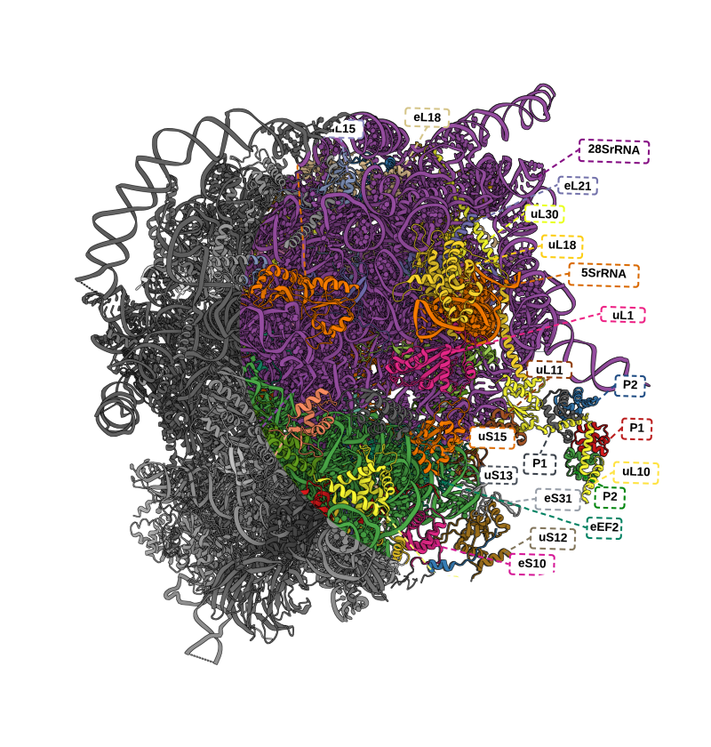
Multiple classes of antibiotics and their binding sites identified on the structure 7K00 (due to Watson et al., 2021) of the E. coli ribosome.

List all unique nonpolymeric ligands along with the structures they bind.

$ curl -X 'GET' \ 
 'https://api.ribosome.xyz/ligands/list_ligands' \ 
 -H 'accept: application/json'

Get residues in the proximity of a given ligand in a structure. Ex. Get all residues within 5 Angstrom of Paromomcyin in E. coli 7K00:

$ curl -X 'GET' \ 
 'https://api.ribosome.xyz/ligands/binding_pocket?source_structure=7K00&chemical_id=PAR&radius=5' \ 
 -H 'accept: application/json'

Attempt to predict a given ligand's binding site in a target structure based on existing site in source structure. Ex. Predict the binding site of PAR(Paromomycin) in E. coli 5AFI using 7K00.PAR as template:

$ curl -X 'GET' \ 
 'https://api.ribosome.xyz/ligands/transpose?source_structure=7K00&target_structure=5AFI&chemical_id=PAR&radius=10'  \ 
  -H 'accept: application/json'

Loci, Functional Sites & Regions of Interest

Coordinates, primary sequence annotations and structural landmarks. Displayed are the Nascent Peptide Exit Tunnel (NPET) geometries produced via the PTC and constriction site landmarks.

Geometry of human ribosome exit tunnel with parts ofuL22uL4eL39proteins displayed.

Geometry of bacterial ribosome tunnel with parts ofuL4uL22uL23proteins displayed. A molecule oferythromycinis present.

The geometry of human mitochondrial ribosome exit tunnel with parts ofuL4muL22muL23mproteins displayed.Nascent chainis traversing the tunnel.

Download a `.ply` file containing the geometry of the human ribosome exit tunnel in 4UG0

$ curl -X 'GET' 'https://api.ribosome.xyz/loci/tunnel_geometry?rcsb_id=4ug0&is_ascii=false' -H 'accept: */*'

The coordinate of the constriction site formed by ribosomal proteins uL4 and uL22 in 8FC4

$ curl -X 'GET' 'https://api.ribosome.xyz/loci/constriction_site?rcsb_id=8fc4'  -H 'accept: application/json'

The coordinate of the peptidyl transferase center (PTC) in the human ribosome 3J7Z

$ curl -X GET https://api.ribosome.xyz/loci/ptc?rcsb_id=3j7z -H 'accept: application/json'

Acknowledgements

We are indebted to the following institutions and projects for being open source or supporting us otherwise

University of British Columbia
Mol* Project
Protein Data Bank
Neo4j Graph Database
HMMER Suite
ChimeraX
Ribovision 2 & The Willaims Lab

How to Cite

@article{kushner2023riboxyz,
  title={RiboXYZ: a comprehensive database for visualizing and analyzing ribosome structures},
  author={Kushner, Artem and Petrov, Anton S and Dao Duc, Khanh},
  journal={Nucleic Acids Research},
  volume={51},
  number={D1},
  pages={D509--D516},
  year={2023},
  publisher={Oxford University Press}
}

Contact

Drop us a line regarding improvements to the service, errors or direct requests for data you couldn't find.